CELL CULTURE SUPERNATE ASSAY
Inter-assay Precision (Precision between assays) Three samples of known concentration were tested in twenty separate assays to assess inter-assay precision. Assays were performed by at least three technicians using two lots of components.To assess linearity of the assay, samples containing and/or spiked with high concentrations of human IL-1β were diluted with the appropriate Calibrator Diluent to produce samples with values within the dynamic range of the assay.This immunoassay is calibrated against highly purified recombinant human IL-1β. The non-WHO reference material for IL-1β 86/552 was evaluated in this kit. The dose response curve of the reference material parallels the Quantikine standard curve. To convert sample values obtained with the Quantikine kit to approximate NIBSC (86/552) units, use the equation below. Cell Culture Supernates – Human peripheral blood mononuclear cells (1 x 10⁶ cells/mL) were cultured in RPMI supplemented with 10% fetal calf serum, 50 μM β-mercaptoethanol, 2 mM L-glutamine, 100 U/mL penicillin, and 100 μg/mL streptomycin sulfate. Cells were stimulated with the agents listed in the table below. Aliquots of the cell culture supernate were removed on days 1, 3, and 5 and assayed for levels of human IL-1β.
Interleukin 6 (IL-6) is a pleiotropic, α-helical, 22-28 kDa phosphorylated and variably glycosylated cytokine that plays important roles in the acute phase reaction, inflammation, hematopoiesis, bone metabolism, and cancer progression (1-5). Mature human IL-6 is 183 amino acids (aa) in length and shares 39% aa sequence identity with mouse and rat IL-6 (6). Alternative splicing generates several isoforms with internal deletions, some of which exhibit antagonistic properties (7-10). Cells known to express IL-6 include CD8+ T cells, fibroblasts, synoviocytes, adipocytes, osteoblasts, megakaryocytes, endothelial cells (under the influence of endothelins), sympathetic neurons, cerebral cortex neurons, adrenal medulla chromaffin cells, retinal pigment cells, mast cells, keratinocytes, Langerhans cells, fetal and adult astrocytes, neutrophils, monocytes, eosinophils, colonic epithelial cells, B1 B cells and pancreatic islet beta cells (2, 11-33). IL-6 production is generally correlated with cell activation and is normally kept in control by glucocorticoids, catecholamines, and secondary sex steroids (2). Normal human circulating IL-6 is in the 1 pg/mL range, with slight elevations during the menstrual cycle, modest elevations in certain cancers, and large elevations after surgery (34-38).
IL-6 induces signaling through a cell surface heterodimeric receptor complex composed of a ligand binding subunit (IL-6 R alpha) and a signal transducing subunit (gp130). IL-6 binds to IL-6 Rα, triggering IL-6 Rα association with gp130 and gp130 dimerization (39). gp130 is also a component of the receptors for CLC, CNTF, CT-1, IL-11, IL-27, LIF, and OSM (40). Soluble forms of IL-6 Rα are generated by both alternative splicing and proteolytic cleavage (5). In a mechanism known as trans-signaling, complexes of soluble IL-6 and IL-6 Rα elicit responses from gp130- expressing cells that lack cell surface IL-6 Rα (5). Trans-signaling enables a wider range of cell types to respond to IL-6, as the expression of gp130 is ubiquitous, while that of IL-6 Rα is predominantly restricted to hepatocytes, monocytes, and resting lymphocytes (2, 5). Soluble splice forms of gp130 block trans-signaling from IL-6/IL-6 Rα but not from other cytokines that use gp130 as a co-receptor (5, 41).
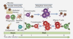
IL-6, along with TNF-α and IL-1, drives the acute inflammatory response. IL-6 is almost solely responsible for fever and the acute phase response in the liver, and it is important in the transition from acute inflammation to either acquired immunity or chronic inflammatory disease (1-5). When dysregulated, it contributes to chronic inflammation in conditions such as obesity, insulin resistance, inflammatory bowel disease, arthritis, and sepsis (2, 5). IL-6 modulates bone resorption and is a major effector of inflammatory joint destruction in rheumatoid arthritis through its promotion of Th17 cell development and activity (1). It contributes to atherosclerotic plaque development and destabilization as well as the development of inflammation-associated carcinogenesis (1, 2). IL-6 can also function as an anti-inflammatory molecule, as in skeletal muscle where it is secreted in response to exercise (2). In addition, it enhances hematopoietic stem cell proliferation and the differentiation of memory B cells and plasma cells (42).
PRINCIPLE OF THE ASSAY
This assay employs the quantitative sandwich enzyme immunoassay technique. A monoclonal antibody specific for human IL-6 has been pre-coated onto a microplate. Standards and samples are pipetted into the wells and any IL-6 present is bound by the immobilized antibody. After washing away any unbound substances, an enzyme-linked polyclonal antibody specific for human IL-6 is added to the wells. Following a wash to remove any unbound antibody-enzyme reagent, a substrate solution is added to the wells and color develops in proportion to the amount of IL-6 bound in the initial step. The color development is stopped and the intensity of the color is measured.This kit is also available in a PharmPak (R&D Systems, Catalog # PD6050). PharmPaks contain sufficient materials to run ELISAs on 50 microplates. Specific vial counts of each component may vary. Please refer to the literature accompanying your order for specific vial counts.


